 |
|
Micro X-ray CT at SPring-8 X-ray CT is the most popular visualization technique. In order to observe the each grains' movement of real sand, we use a micro X-ray CT system at SPring-8. |
|
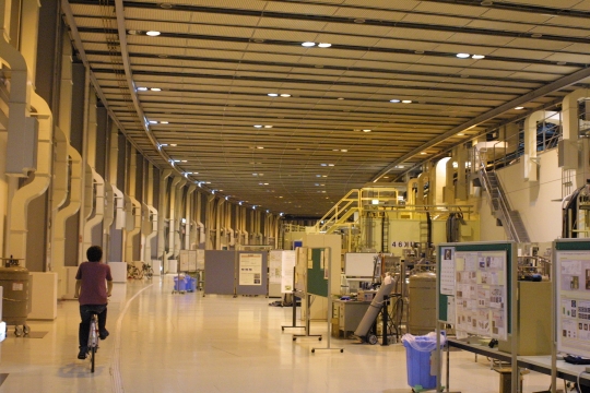
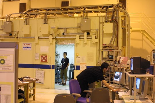
[1] What is SPring-8 ? SPring-8, the world's largest third-generation synchrotron radiation facility, has laboratories, BL20B2 and BL47XU, at which a well designed micro x-ray system is available. Its high flux density x-ray beam is naturally well collimated and monochromatic, which enables us a very high spatial resolution of x-ray CT (around 10 m and 1 m at BL20B2 and BL47XU, respectively). 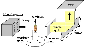
Figure 1 X-ray CT system in SPring-8 to SPring-8 web site [2] CT reconstruction Using a special image processing technique, 3-D shape of each grain in a specimen is quantified. 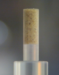
Figure 2 Example of specimen (Toyoura sand) 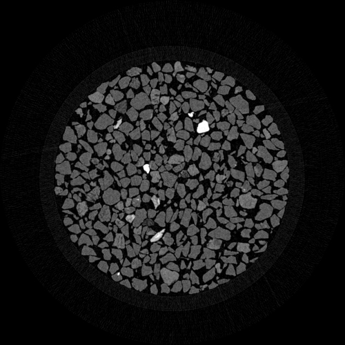
Figure 3 Example of CT reconstructed image (plan view) example of reconstructed 3D image of a grain (avi) [3] Micro triaxial test In order to observe 3D grain motion inside the triaxial specimen, we developed a micro-triaxial test apparatus. The resulting image was quite clear and we can see the motion of the grains. 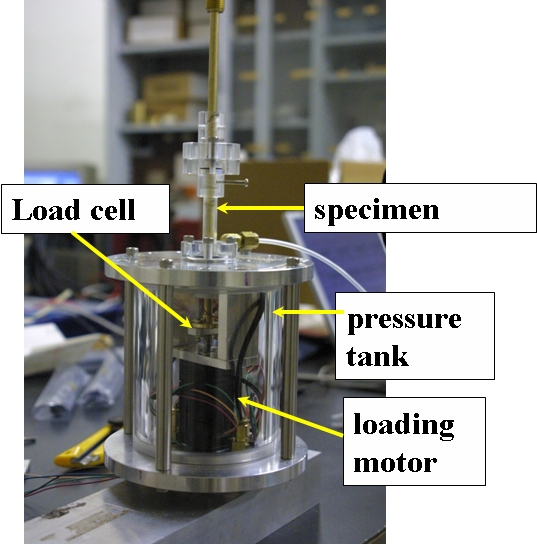
Figure 4 Micro triaxial test apparatus 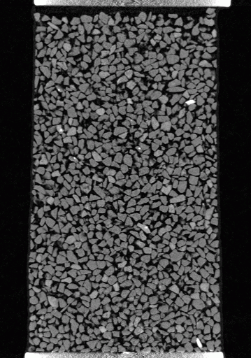 Figure 5 graion motion within a vertical cross section of a triaxial specimen (gif animation) [3] SPring-8 user experimental reports 2003A0127 2003B0479 2004A0165 2004A0163 
 |
